Ovarian Cyst Pelvic Ultrasound Female / Benign And Malignant Tumours Of The Female Reproductive System Undergraduate Diagnostic Imaging Fundamentals / A pelvic ultrasound allows quick visualization of the female pelvic organs and structures including the uterus, cervix, vagina, fallopian tubes and ovaries.
Ovarian Cyst Pelvic Ultrasound Female / Benign And Malignant Tumours Of The Female Reproductive System Undergraduate Diagnostic Imaging Fundamentals / A pelvic ultrasound allows quick visualization of the female pelvic organs and structures including the uterus, cervix, vagina, fallopian tubes and ovaries.. The most common indications for imaging of the pelvis in girls include ambiguous genitalia, prepubertal bleeding, primary amenorrhea, pelvic mass, and pelvic pain. A 51 year old, gravida 3 para 2, comes to the office because a routine health maintenance examination. These are called simple ovarian cysts. Pelvic ultrasound shows a simple right ovarian cyst measuring 4 cm in diameter. Her last menstrual period was at age 49.
Ovarian cysts, also known as ovarian masses or adnexal masses, are frequently found incidentally in asymptomatic women. Ultrasound imaging of the pelvic area is used to help determine the cause of symptoms such as pelvic pain and abnormal bleeding in women. She has no specific complaints at this visit. This is called a tubal pregnancy. The left ovary is mostly displaced by a large 7.5cm ovarian cyst.
Torsion Of Huge Dermoid Cyst In Adolescent Girl A Case Report Sciencedirect from ars.els-cdn.com These scans often also find cysts in the ovaries. It is the single most effective way of imaging and characterizing ovarian cysts. Follicle production is normal in all premenopausal women. It is excellent at this task as the ovaries naturally contain fluid called follicles or cysts. Eggs (ova), which develop and mature in the ovaries, are released in monthly cycles during the childbearing years. The left ovary is mostly displaced by a large 7.5cm ovarian cyst. There were no ovarian cancers found. Most of the time those extra exams are not needed.
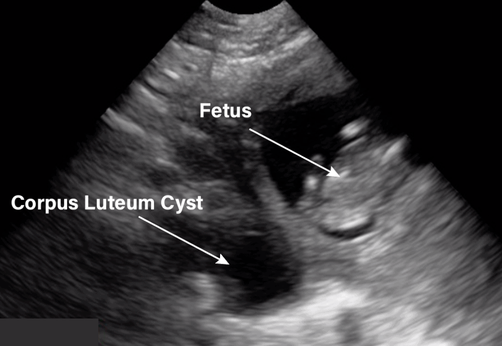
In premenopausal women, a dominant cyst, or corpus luteum, forms with each cycle.
A 51 year old, gravida 3 para 2, comes to the office because a routine health maintenance examination. If the ovarian cyst does not enlarge or if it resolves during the period of watchful waiting, it does not usually require surgical removal. A pelvic ultrasound test is done to: Check for growths or masses like ovarian cysts or uterine fibroids. The right ovary measures up to 3.4cm and has a normal ultrasound appearance. Ultrasound is the first line among medical diagnostic radiology imaging tools to diagnose ovarian cancer. Women have two ovaries — each about the size and shape of an almond — on each side of the uterus. Premenopausal women — in premenopausal women, watchful waiting usually involves monitoring for symptoms (pelvic pain or pressure) and repeating the pelvic ultrasound after six to eight weeks. Find the cause of urinary problems. These scans often also find cysts in the ovaries. The most common indications for imaging of the pelvis in girls include ambiguous genitalia, prepubertal bleeding, primary amenorrhea, pelvic mass, and pelvic pain. Ovarian cysts in teenagers are fairly common and typically don't cause further issues. What are ovarian cysts & pelvic masses ovarian cysts are a common occurrence in countless women and often resolve on their own.
Ultrasound uterine wt 0.16 0.80 < 0.001 ≥ 200 g patient wt 0.09 0.73. Pelvic ultrasound shows a simple right ovarian cyst measuring 4 cm in diameter. A retrospective review of an entire clinical series of 152 women over 50 years of age, in whom cystic lesions without solid parts had been diagnosed by ultrasound, found there were no malignancies in 58 completely anechoic lesions less than 5 cm in diameter. Ovarian cysts in teenagers are fairly common and typically don't cause further issues. These are called simple ovarian cysts.
Gynecology Pelvic Ultrasound Made Easy Step By Step Guide Pocus 101 from pocus101.b-cdn.net Ovarian cysts in teenagers are fairly common and typically don't cause further issues. Follicle production is normal in all premenopausal women. A cyst on your ovary can be found during a pelvic exam. All females of reproductive age will likely develop functional cysts at some point in their life. It is excellent at this task as the ovaries naturally contain fluid called follicles or cysts. Ultrasound is the first line among medical diagnostic radiology imaging tools to diagnose ovarian cancer. One well done study (jermy, 2001), looked at the reliability of ultrasound to make a correct diagnosis for possible endometriosis or dermoid types of complex ovarian cysts. For example, limited field of view, obscuration of pelvic organs by the presence of bowel gas, dependency on patient size, and dependency on the skill and experience of the operator.
There is no marked free fluid within the pelvis.
If the ovarian cyst does not enlarge or if it resolves during the period of watchful waiting, it does not usually require surgical removal. Ultrasound uses a transducer that sends out ultrasound waves at a frequency too high to be heard. These cysts are well evaluated by ultrasound. On bimanual examination, a small, right adnexal mass is palpated. See if a fertilized egg is growing outside the uterus. Her last menstrual period was at age 49. The most common indications for imaging of the pelvis in girls include ambiguous genitalia, prepubertal bleeding, primary amenorrhea, pelvic mass, and pelvic pain. Transabdominal pelvic ultrasound can detect most larger abnormalities such as large fibroids, ovarian cysts, neoplasms, etc. In general for pocus exams, it is usually good to start with the transabdominal ultrasound and then use the transvaginal approach if needed. Of 10 small lesions (less than 5 cm in di … An ultrasound review of pelvic pathology judi m bender md january 20, 2016. Depending on its size and whether it's fluid filled, solid or mixed, your doctor likely will recommend tests to determine its type and whether you need treatment. All females of reproductive age will likely develop functional cysts at some point in their life.
All females of reproductive age will likely develop functional cysts at some point in their life. Hemorrhagic cysts septations with internal echoes and no flow. Follicle production is normal in all premenopausal women. See if a fertilized egg is growing outside the uterus. These scans often also find cysts in the ovaries.
Hot Tips Locating The Ovaries On Transabdominal Ultrasound Youtube from i.ytimg.com These cysts are well evaluated by ultrasound. Ultrasound uterine wt 0.16 0.80 < 0.001 ≥ 200 g patient wt 0.09 0.73. Ultrasound uses a transducer that sends out ultrasound waves at a frequency too high to be heard. After the mass was removed it was found that ultrasound was successful in predicting 96% of endometriosis cysts and 97% of dermoids. There were no ovarian cancers found. All females of reproductive age will likely develop functional cysts at some point in their life. A cyst can form during ovulation when the follicle that's supposed to rupture and release an egg fails to do so. There is no marked free fluid within the pelvis.
The right ovary measures up to 3.4cm and has a normal ultrasound appearance.
Most cysts are harmless, but some may cause problems such as rupturing, bleeding, or pain; A cyst on your ovary can be found during a pelvic exam. If your healthcare provider finds an unexpected cyst or enlarged ovary during a pelvic exam, you should have a vaginal ultrasound to assess for cancer. Another type of cyst happens when fluid accumulates in the follicle. See if a fertilized egg is growing outside the uterus. The left ovary is mostly displaced by a large 7.5cm ovarian cyst. Followup of ovarian cysts in premenopausal women •simple cysts •1.<3cm=normal finding •2. Check for growths or masses like ovarian cysts or uterine fibroids. Because of concerns about ovarian cancer, these cysts may be followed with repeated imaging over many years. Find the cause of urinary problems. Ovarian cysts in teenagers are fairly common and typically don't cause further issues. A pelvic ultrasound allows quick visualization of the female pelvic organs and structures including the uterus, cervix, vagina, fallopian tubes and ovaries. What are ovarian cysts & pelvic masses ovarian cysts are a common occurrence in countless women and often resolve on their own.
Follicle production is normal in all premenopausal women pelvic ultrasound female. An ultrasound review of pelvic pathology judi m bender md january 20, 2016.


0 Komentar